Deciphering cortical circuit alterations in neurodegenerative diseases
To investigate neurodegeneration-related circuit dysfunction, we employ in vivo two-photon imaging of neuronal activity in awake behaving mice.
Chronic imaging in the R6/2 mouse model of Huntington’s disease revealed an increase in activity in the primary motor cortex, which preceded the onset of motor impairments in the mice (Fig. 1) (Burgold et al. 2019).
Proteomic and histological analyses suggested that these alterations in the cortical network might be due to inhibitory synapse defects (Fig. 2).
Building upon these findings, we are now investigating the involvement of cortical GABAergic interneuron types in Huntington’s disease.
We combine imaging with behavioral tracking in order to reveal neuronal correlates of motor defects (Fig. 3).
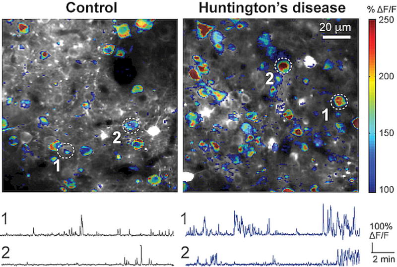
Figure 1.
Activity maps of imaged areas in the primary motor cortex of control and R6/2 Huntington’s disease mice (top) and example calcium traces of single neurons (bottom). Adapted from Burgold et al., 2019
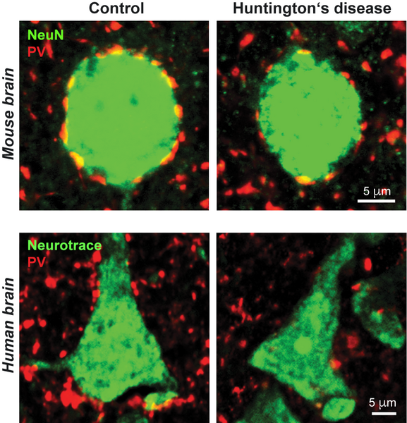
Figure 2.
Perisomatic GABAergic inhibitory synapses from parvalbumin (PV) interneurons (red) on the cell bodies of cortical pyramidal cells (green) in the primary motor cortex of R6/2 mice (top) and Huntington’s disease patients (bottom). Note the reduction in PV synapses in Huntington’s disease mice and patients. Adapted from Burgold et al., 2019.
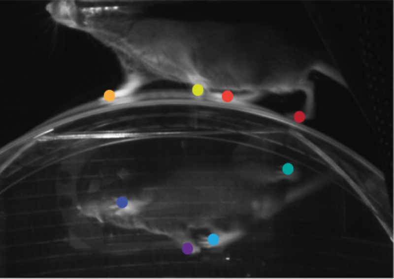
Figure 3.
Three-dimensional tracking of motor behavior during in vivo imaging in head-fixed mice. Colored dots indicate deep learning-assisted paw tracking.