Neuromorphology
Neuromorphology
Morphology and neurochemistry of the amygdala
Affective disorders such as depression, anxiety syndromes and posttraumatic stress disorder are hitherto incompletely understood, frequent disorders with severe consequences for the individuum and society. The corpus amygdaloideum (amygdala) is the telencephalic center for processing of emotional, particularly fear- and anxiety-inducing stimuli. Malfunctions in interconnections of this brain area may contribute to affective disorders. We analyze the structure of amygdaloid networks and investigate which factors could be responsible for normal function and pathological changes, focusing on the role of monoaminergic afferents and peptidergic systems of the amygdala.
In particular, we are focusing on the following questions:
- Which are the target neurons of dopaminergic or serotonergic afferents in different amygdala regions?
- Which receptors or receptor combinations mediate the effect of monoaminergic innervation on identified target neurons in the amygdala?
- Which interactions are present between monoaminergic afferents and specific anxiogenic/anxiolytic peptidergic systems in different amygdala regions?
- Does stress experience evoke morphological and/or neurochemical alterations in the amygdala?
- What alterations are found in monoamínergic and peptidergic systems in the amygdala in animal models for affective disorders in the human, and how do possible alterations correlate with altered behavior?
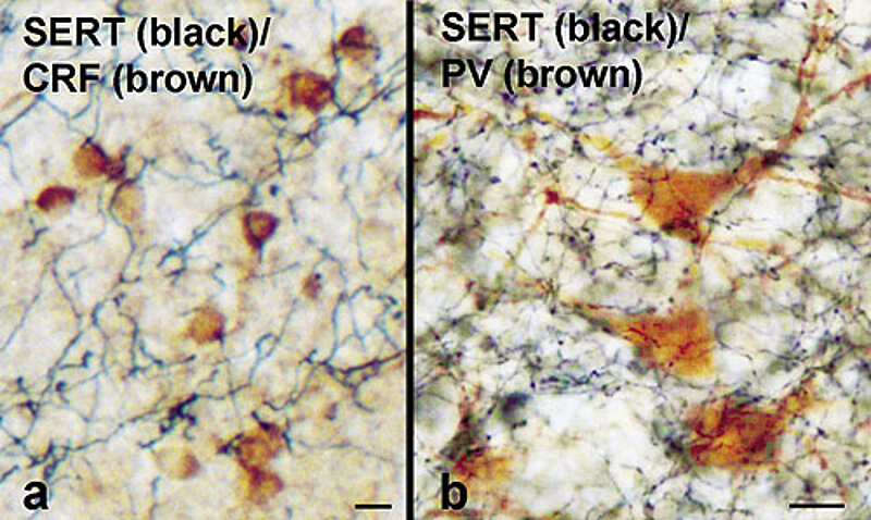
Fig. 1: Corticotropin-releasing factor (CRF;a)-, und Parvalbumin (PV;b)-produzierende Neuronen in der Amygdala werden von serotonergen Axonen kontaktiert. Doppel-Immunreaktionen, Ratte.
Maßbalken: 10 µm
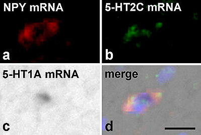
Fig. 2: Einzelne Neuropeptid Y(NPY) mRNA produzierende Neurone (a) in der Amygdala coexprimieren mRNA für die beiden Serotonin-Rezeptor Subtypen 5-HT2C (b) und 5-HT1A (c). (d) Überlagerung der Reaktionen in (a-c) Dreifach-in situ Hybridisierung, Ratte.
Maßbalken: 20 µm
Selected Publications:
Asan E, Steinke M, Lesch KP (2013)
Serotonergic innervation of the amygdala: targets, receptors, and implications for stress and anxiety.
Histochem Cell Biol 139:785-813.
Bonn M, Schmitt A, Lesch KP, Van Bockstaele EJ, Asan E (2013).
Serotonergic innervation and serotonin receptor expression of NPY-producing neurons in the rat lateral and basolateral amygdaloid nuclei.
Brain Struct Funct 218:421-35.
Bonn M, Schmitt A, Asan E (2012).
Double and triple in situ hybridization for coexpression studies: combined fluorescent and chromogenic detection of neuropeptide Y (NPY) and serotonin receptor subtype mRNAs expressed at different abundance levels.
Histochem Cell Biol. 137:11-24.
Nietzer SL*, Bonn M*, Jansen F, Heiming RS, Lewejohann L, Sachser N, Asan ES*, Lesch KP*, Schmitt AG* (2011).
Serotonin transporter knockout and repeated social defeat stress: impact on neuronal morphology and plasticity in limbic brain areas.
Behav Brain Res 220:42-54.
Asan E, Yilmazer-Hanke DM, Eliava M, Hantsch M, Lesch KP, Schmitt A (2005).
The corticotropin-releasing factor (CRF)-system and monoaminergic afferents in the central amygdala: investigations in different mouse strains and comparison with the rat.
Neuroscience 131:953-67
Eliava M, Yilmazer-Hanke D, Asan E (2003).
Interrelations between monoaminergic afferents and corticotropin-releasing factor-immunoreactive neurons in the rat central amygdaloid nucleus: ultrastructural evidence for dopaminergic control of amygdaloid stress systems.
Histochem Cell Biol. 120:183-97.
Asan E (1998)
The catecholaminergic innervation of the rat amygdala.
Adv Anat Embryol Cell Biol. 142:1-118.
Asan E (1997)
Ultrastructural features of tyrosine-hydroxylase-immunoreactive afferents and their targets in the rat amygdala.
Cell Tissue Res 288:449-69
Asan E (1997)
Interrelationships between tyrosine hydroxylase-immunoreactive dopaminergic afferents and somatostatinergic neurons in the rat central amygdaloid nucleus.
Histochem Cell Biol 107(1):65-79
Asan E (1995)
The adrenergic innervation of the rat central amygdaloid nucleus: a light and electron microscopic immunocytochemical study using phenylethanolamine N-methyltransferase as a marker.
Anat Embryol 192:471-81.